© Center for Endometriosis Care/Ken Sinervo MD, MSc, FRCSC. All rights reserved. No reproduction permitted without written permission. Edition current as of 2025. No external funding was utilized in the creation of this material. The Center for Endometriosis Care neither endorses nor has affiliation with any resources cited herein. The following material is for informational purposes only and does not constitute medical advice.
Gut Check: Why the Appendix Matters in Endometriosis Care
Endometriosis has long been defined as a systemic, inflammatory disease characterized by the presence of ‘endometrium-like tissue’ found outside the uterus (Collins, 1951; Uohara et al., 1975; Jansen et. al., 1986; Hwang et al., 2016; International Working Group, 2021; Taylor et al., 2021; Akano et al., 2025). More recently referred to as a spectrum disease with a variety of subtypes and clinical presentations, endometriosis is routinely marked by pain (often chronic), inflammation, infertility, organ dysfunction, and much more (International Working Group, 2021; Mariadas et al., 2025). Alterations in certain biological processes of the endocrine and immune systems have been observed with the disease (Herington et al., 2011), and it is embodied by a complexity of multiple immunologic abnormalities, endocrine alterations, and the unusual expression of adhesion molecules (Kyama et al., 2008). In the simplest of terms: endometriosis is an inflammatory disease wherein tissue somewhat resembling the native endometrium is found elsewhere in the body, triggering a cascade of various reactions and symptoms.
Despite its age-old tenuous relationship to the lining of the uterus, endometriosis is no longer regarded as merely a gynecologic condition or 'just a bad period;’ acknowledging the full scope of its impact is essential to delivering better outcomes for all those affected by the disease. To that end, it is important to recognize that endometriosis has been documented in virtually every organ system including a variety of extragenital sites, such as the appendix.
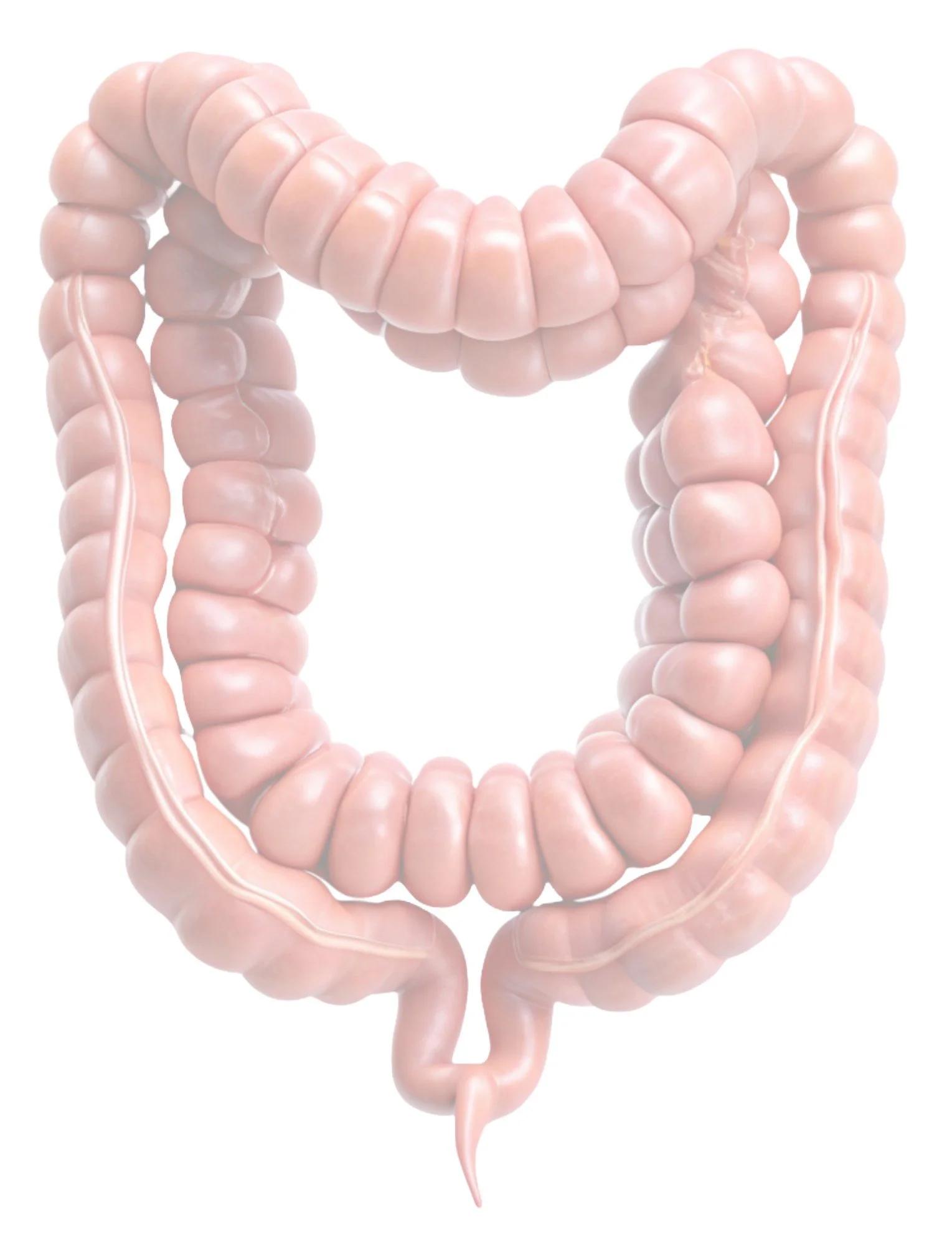
The appendix is a small, finger-like structure that protrudes from the colon on the lower right side of the abdomen. Current research suggests it may play a role in gut health and the immune system, though it is not essential for survival. Most people recognize appendicitis as a well-known medical concern; what’s less known is that the appendix can also be affected by endometriosis and other conditions, which may not show up on tests or cause typical symptoms in those affected.
Far from a recent discovery, appendiceal endometriosis has been routinely documented in the ‘modern’ literature since for more than a century (e.g., Dougal, who described the case of an "appendicular adenomyoma" in 1923; Outerbridge, 1925; Seelig, 1926; Cattell et al., 1936; Cotte et al., 1946; Shemilt, who reported an endometrioma of the caecum that was found to be obstructing the lumen of the appendix in 1949; and many others). In 1951, Collins identified more than 100 cases that had been published from 1860-1951, while Romanus (1952) later suggested that the true incidence within that time frame was under-reported, noting that Collins had overlooked at least 11 additional reports in his review. These and other early observations underscore that appendiceal involvement in endometriosis is far from a new phenomenon, but one that remains underrecognized in both clinical practice and research still today.
Collins (1951) historically noted that endometriosis of the appendix was becoming more frequently recognized even more than 70 years ago because the pathologist and the surgeon were increasingly learning to identify its gross and microscopic characteristics. Historically considered a manifestation of (then so-called) ‘external endometriosis,’ Sampson’s widely-cited theory of retrograde menstruation (1921) was initially used to explain all forms of the disease at the time, including appendix-involved cases. However, as Collins and others argued even then, Sampson's Theory could not - and still does not - account for the numerous documented cases of endometriosis including and especially in distant and anatomically unrelated sites; i.e., the lungs (Thompson, 1926; Bungeler & Fleury Silveira, 1939), the thigh (Mankin, 1933; Schlicke, 1946), the extensor carpi radialis muscle of the forearm (Navratil & Kramer, 1936), and many others. Later, countless studies including by Hayata et al. (1991) determined it was ‘unlikely that endometriosis is wholly derived from the native endometrium.’ Indeed, in his own early case studies, Sutton (1952) asserted that the 'explanation of the disease within the wall of the appendix’ was best left ‘to the imagination of the reader.’ Such findings have long highlighted the limitations of Sampson's etiologic explanation, pointing instead to the multifactorial pathogenesis underlying endometriosis and variety of mechanisms driving the disease.
Appendiceal endometriosis may cause no symptoms at all but when present, can closely resemble other conditions. Symptoms may mimic acute or chronic appendicitis, perforation, or intussusception, and can include right lower quadrant pain, rebound tenderness, bloating, diarrhea, constipation, rectal pain, nausea, vomiting, signs of an obstruction, and more. Symptoms and pain can be cyclical and coincide with or worsen around periods in those who menstruate, but may also occur in non-cyclical, nonspecific patterns. However, there are cases where pain does not correlate with the menstrual cycle (Kawaguchi et al., 2024) and it can present in varied and nonspecific ways. For example, of the 12 patients reported in a 1975 case series by Uohara et al., none displayed symptoms of acute appendicitis. 13 of the 15 cases presented by Sutton & Hardy in 1952 also had no gastrointestinal symptoms at all. The disease can also be found in those with no prior diagnosis of endometriosis (Mehrotra et al., 2025), and given its wide variety of presentations, the gold standard for a definitive diagnosis is histopathological examination of the excised appendix (Lee et al., 2024).
While reports vary today, involvement of the gastrointestinal tract with endometriosis generally is reported to affect between 3-37% of patients with the disease; when it does, the rectosigmoid (72%), rectovaginal septum (13%), small intestine (7%), cecum (3.6%) and appendix (3%) are most commonly involved (Kawaguchi et al., 2024; Gurluler et al., 2025). More recently, Bolcatto et al. (2025) estimated that appendiceal endometriosis itself ranges from 0.05 %-1.7 % overall in patients with the disease. Other reports indicate the prevalence of appendiceal endometriosis is approximately 2.8% (Centini et al., 2024) and in those patients affected by endometriosis of the appendix, concomitant ovarian and/or bladder endometriosis are more frequently encountered. Schrempf et al. (2024) also reported that the incidence of appendiceal endometriosis among patients undergoing appendectomy for suspected appendicitis is even higher than previously estimated in autopsy studies and cohorts with biopsy-proven endometriosis, and a markedly increased likelihood of appendiceal endometriosis occurs in patients diagnosed with diaphragmatic endometriosis (Pagano et al., 2024). Significant association has also been reported by Centini et al. (2024) between appendiceal, ovarian, and bladder endometriosis.
Appendix-associated endometriosis has also been outlined in multiple case reports demonstrating challenges during pregnancy, even as far back as the late 1940s (Bogatko, 1949; Faucheron et al., 2008; Stiegler et al., 2024; Arachchilage et al., 2025). Other very rare complications can also occur, such as appendiceal intussusception. Intussusception happens when the appendix segment is pulled into itself or into the cecum (the beginning of the large intestine). While considered distinctly uncommon, the condition can cause a blockage (Milošević et al., 2025) and other potentially serious outcomes ranging from ischemia to increased risk of perforation. Though intussusception occurs due to a myriad of causes, the literature contains several case reports of the condition being specifically caused by endometriosis; e.g., the first recorded report by Deacon (1949), in which his patient had no prior diagnosis of endometriosis; a 21 year old patient in whom the intussusception was missed on both CT scan and Laparoscopy (El Sadaney et al., 2024); an older patient who had intermittent chronic abdominal pain that did not coincide with menses and no prior diagnosis of endometriosis (Kawaguchi et al., 2024); and the Kosaka et al. case (2025) of appendiceal intussusception with cecal involvement caused by endometriosis histologically confirmed at the appendiceal tip. The latter encouraged the authors to advise colleagues that endometriosis, while an uncommon cause of intussusception, should be considered among the differential diagnoses.
Though considered less common than other forms of the disease, endometriosis of the appendix is nevertheless clinically significant when it does occur, and importantly, Guo et al. (2024) note that even a “macroscopically normal looking appendix may harbor endometriosis.” Some authors have even determined that among their patient populations, terminal ileum/appendix endometriosis was the “most common extra-pelvic cause of acute bowel obstruction” (Gurluler et al., 2025). Other have concluded that when the appendix is “not considered in endometriosis pathology, patients are at increased risk for unresolved pain and further laparoscopic procedures” (Klein et al, 2023).
Centini et al. (2024) noted that given the prevalence of appendiceal involvement, counseling regarding the potential need for appendectomy during endometriosis surgery should be considered, adding to a growing body of evidence that suggests removing the appendix during surgery for endometriosis can prevent future issues and reduce diagnostic uncertainty and that surgical assessment of the organ is critical (Calcagnotto et al., 2024). However, the suggestion of prophylactic/preventive appendectomy being performed during gynecologic surgery is not new (Krone et al., 1989). Even in the 1950s, researchers concluded that 'in patients with pelvic endometriosis, involvement of the vermiform appendix is not uncommon; it would therefore seem advisable, when surgery for pelvic endometriosis is undertaken, to perform incidental appendectomy' (Sutton et al., 1952). Our Center’s own Wendy Winer suggested nearly 25 years ago (Lyons, Winer, Woo, 2001) that those patients undergoing laparoscopic surgery should be counseled on the high frequency of abnormal findings in the appendix, including endometriosis. Other data have since concluded that laparoscopic appendectomy can and should be performed at time of excision of Stage IV endometriosis and be routinely considered in the subset of Stage IV endometriosis patients with colorectal involvement in particular (Guo et al., 2023).
Of course, the decision to remove the appendix during endometriosis surgery depends on the individual case, but evidence suggests it can be valuable, especially when there is visible or suspected endometriosis or other condition(s) involving the appendix; when the patient reports chronic or cyclical right lower quadrant pain without another clear source; if the appendix looks scarred, thickened or is otherwise abnormal appearing; when there is a history of appendicitis-like symptoms, even in absence of prior diagnosis; in cases of deeply infiltrating endometriosis where multi-organ involvement is likely; when the appendix is involved by adhesions or anatomic distortion; if the patient has had incomplete relief from prior surgeries or presents with persistent GI complaints; and/or as a preventive measure given the high incidence of pathology even in otherwise normal looking appendices. Prophylactic appendectomy appears to be a worthwhile consideration in patients with chronic pelvic pain overall as well, given the high frequency of pathological findings that may be revealed following appendectomy as Winer et al. noted years ago. The literature documents a range of conditions discovered upon histologic examination, including acute and chronic appendicitis, hyperplastic polyps, follicular hyperplasia, even malignancies such as well-differentiated adenocarcinoma or mucinous cystadenoma and many others. One recent study revealed that even as many as 52.9% of cases found abnormal pathological findings during gynecologic surgeries in which appendectomy was performed (Yagur et al., 2024).
As a result, it is widely accepted in the Minimally Invasive Gynecologic Surgery community that removal of the appendix in patients with endometriosis contributes to optimal surgical management, while relying on abnormal appendiceal appearance for removal may leave endometriosis-affected appendices behind to cause ongoing pain and symptoms (Nikou et al., 2021). Taken in full context, a century of evidence-based literature clearly outlines the possibility of appendix-involved endometriosis and provides abundant evidence in support performing an appendectomy at the time of surgery.
Although appendiceal endometriosis is considered less common than other forms of the disease, it remains a clinically significant entity that should not be overlooked. Its potential to mimic other conditions, contribute to persistent or unexplained symptoms, and coexist with other pelvic or gastrointestinal pathologies underscores the importance of thorough evaluation during endometriosis care. Incorporating appendectomy, especially when abnormalities are present or symptoms warrant, can enhance diagnostic accuracy, support comprehensive care, and reduce the likelihood of missed disease. In our Center, it is considered part of our integrated surgical management that helps us identify occult disease, prevent future complications, and reduce the need for repeat procedures - particularly in those with chronic pain and/or gastrointestinal symptoms. This proactive approach reflects our commitment to addressing all potential sources of pain and advancing long-term outcomes for our patients.
We have long recognized the role the appendix can play in persistent or unexplained symptoms, and more than a century of evidence-based literature supports what our team has previously reported: the possibility of appendix-involved endometriosis and the importance of considering appendectomy during endometriosis surgery when clinically appropriate. Because appendiceal endometriosis can mimic other conditions, contribute to ongoing symptoms, and/or coexist with other pelvic or gastrointestinal issues, thorough evaluation is critical. It can easily be overlooked, which is why we work so closely with our interdisciplinary team of radiologists, GI specialists and others - to ensure all potential sources of pain are identified and addressed through our truly collaborative approach to care.
Akano OP, Amos VO, Fawole IO, Oyindamola AC, Ajayi AF. Epigenetics' Responsibility in Endometriosis: A comprehensive assessment. J Steroid Biochem Mol Biol. Published online June 11, 2025.
Arachchilage CE, Stevens S, van der Hock S, Richardson A. Appendiceal Endometriosis Causing Acute Appendicitis in Pregnancy. Archives of Clinical and Medical Case Reports. 9 (2025): 53-55.
Bogatko FL. Endometriosis of the appendix in conjunction with pregnancy. J Am Med Womens Assoc. 1949;4(6):230-232.
Bolcatto A, Erina M, Mandojana FI, Bruera N, Doniquian AM, Viscido GR. Acute appendicitis due to appendiceal endometriosis: Two case report and literature review. Int J Surg Case Rep. 2025;126:110743.
Bungeler W, Fleury Silveira D. Consideracoes sobre a patogenia das endometrioses (a proposito de 3 casos de endometriose externa). Arg. Cir. Clin. E exper. 3:169-171 (June) 1939.
Calcagnotto H, Rostirolla G, Michelon A, Michelon E, Brambilla, E. (2024) Surgical Management of Appendicular Endometriosis: A Series of Cases. Open Journal of Obstetrics and Gynecology, 14, 1867-1872.
Cattell RB, Swinton NW. Endometriosis: with particular reference to conservative treatment. N Engl J Med 1936;214:341-6.
Centini G, Ginetti A, Colombi I, et al. Endometriosis of the appendix: prevalence, associated lesions, and proposal of pathogenetic hypotheses. A retrospective cohort study with prospectively collected data. Arch Gynecol Obstet. 2024;310(3):1669-1675.
Collins DC. Endometriosis of the vermiform appendix; review of literature, with addition of nine new instances, one of which caused severe melena. AMA Arch Surg. 1951;63(5):617-622.
Cotte G, Sournia J. L'endometriose de l'appendice [Endometriosis of the appendix]. Rev Fr Gynecol Obstet. 1946;41:24-31.
Deacon AL. Endometriosis and intussusception of the appendix. J Obstet Gynaecol Br Emp. 1949;56(4):586-590.
Dougal D. Adenomyoma involving the Vermiform Appendix. BJOG (1923): An International Journal of Obstetrics & Gynaecology, 30: 224-225.
Faucheron JL, Pasquier D, Voirin D. Endometriosis of the vermiform appendix as an exceptional cause of acute perforated appendicitis during pregnancy. Colorectal Dis. 2008;10(5):518-519.
Galaviz VD, Nguyen AD, Sticco PL, Downing KT. Appendectomy in endometriosis: an update on surgical indications and management of uncommon diseases. Curr Opin Obstet Gynecol. 2023;35(4):377-382.
Guo C, Chen MZ, Chiu T, Condous G, Barto W. The appendix in endometriosis. Aust N Z J Obstet Gynaecol. 2023;63(6):792-796.
Gurluler E, Isik O, Ugras N, Sahin A, Sen M, Yilmazlar T. Intestinal endometriosis amongst other extra-pelvic endometriosis foci presenting as acute/subacute bowel obstruction in women of reproductive age: a retrospective case series study. BMC Surg. 2025;25(1):12.
Hayata T, Matsu T, Kawano Y, Matsui N, Miyakawa I. Scanning electron microscopy of endometriotic lesions in the pelvic peritoneum and the histogenesis of endometriosis. Int J Gynaecol Obstet. 1992;39(4):311-319.
Herington JL, Bruner-Tran KL, Lucas JA, Osteen KG. Immune interactions in endometriosis. Expert Rev Clin Immunol. 2011;7(5):611-626.
Hwang MS, Kim YD, Shin SY, et al. Korean J Gastroenterol. 2016;68(4):214-217.
International Working Group of AAGL, ESGE, ESHRE and WES, Tomassetti C, Johnson NP, et al. An international terminology for endometriosis, 2021. Hum Reprod Open. 2021;2021(4):hoab029.
Jansen RP, Russell P. Nonpigmented endometriosis: clinical, laparoscopic, and pathologic definition. Am J Obstet Gynecol. 1986;155(6):1154-1159.
Kawaguchi Y, Maekawa K, Hashimoto T, et al. Intussusception of the appendix secondary to endometriosis: a case report. Surg Case Rep. 2024;10(1):261.
Klein S, Tourangeau-Young R, Biglione A. Appendiceal Endometriosis Presenting As Chronic Appendicitis: A Case Report. Cureus. 2023;15(4):e37825.
Kosaka S, Hirokawa T, Miyai H, et al. Minimum Invasive Surgery for Appendiceal Intussusception Caused by Isolated Endometriosis; Surgical Technique for an Unreturned Case. Asian J Endosc Surg. 2025;18(1):e70059.
Krone HA, Sperke E. Die prophylaktische Appendektomie bei gynäkologischen Eingriffen. Bericht über 1718 Fälle [Preventive appendectomy in gynecologic surgery. Report of 1,718 cases]. Geburtshilfe Frauenheilkd. 1989;49(12):1035-1038.
Kyama CM, Overbergh L, Mihalyi A, et al. Endometrial and peritoneal expression of aromatase, cytokines, and adhesion factors in women with endometriosis. Fertil Steril. 2008;89(2):301-310.
Lee RS, Hemida Y, James D. Appendiceal Endometriosis: A Rare Case of Endometriosis Mimicking Acute Appendicitis. Cureus. 2025;17(3):e81280. Li W, Chen Y, Hong X, Zhang S. Endoscopic management of appendiceal endometriosis. Gastrointest Endosc. 2025;101(2):476-477.
Lyons TL, Winer WK, Woo A. Appendectomy in patients undergoing laparoscopic surgery for pelvic pain. J Am Assoc Gynecol Laparosc. 2001;8(4):542-544.
Mankin ZW. Beitrage zur Lehre von den Endometrioiden Heterotopien. Arch. Klin. Chir. 175:314-334, 1933.
Mariadas H, Chen JH, Chen KH. The Molecular and Cellular Mechanisms of Endometriosis: From Basic Pathophysiology to Clinical Implications. Int J Mol Sci. 2025;26(6):2458.
Mehrotra N, Trovato A, Massier C. Small Bowel Obstruction Secondary to Endometriosis Affecting the Appendix in a Patient With a Virgin Abdomen. Cureus. 2025;17(2):e79717.
Milošević S, Bubanja M, Zugic A, et al. Appendiceal Intussusception: A Rare Diagnosis and the Role of Imaging in Its Detection. Diagnostics (Basel). 2025;15(3):256.)
Navratil E, Kramer A. Endometriose in der Armmuskulatur. Klinische Wochenschrift. 1936 Nov;15:1765-70.
Nikou AF, Tenzel NS, Hua P, Orbuch L, Orbuch IK. Appendectomy Should Be Performed During Minimally Invasive Surgery for Endometriosis. JSLS. 2021;25(1):e2020.00095.
El Sadaney A, Sandhu AD, Parvinian A, Nehra A, Suman G. Appendiceal intussusception due to endometriosis presenting as acute right lower quadrant pain. BJR Case Rep. 2024;10(5):uaae032.
Outerbridge GW. Cystic Lesions of possible Endometrial Origin in the Appendix. Am. J. Obst. & Gynec. 10:545, 1925.
Pagano F, Dedes I, Imboden S, Mueller MD. Connecting the dots: Exploring appendiceal endometriosis in women with diaphragmatic endometriosis. Eur J Obstet Gynecol Reprod Biol. 2024;302:134-140.
Papadia S, Bellone F. Sulle Inclusioni Ghiandolari Endometrio-Simili Nei Linfonodi Pelvici [On Endometrium-Like Lymph Node Inclusions In The Pelvic Lymph Nodes]. Minerva Ginecol. 1964;16:223-227.
Romanus R. Endometriosis of the vermiform appendix. AMA Arch Surg. 1952;65(5):682-683.
Sampson JA. Perforating Hemorrhagic (Chocolate) Cysts Of The Ovary: Their Importance And Especially Their Relation To Pelvic Adenomas Of Endometrial Type ("Adenomyoma" Of The Uterus, Rectovaginal Septum, Sigmoid, Etc.). Arch Surg. 1921;3(2):245–323.
Sampson, JA. The life history of ovarian hematomas (hemorrhagic cysts) of endometrial (müllerian) type. American Journal of Obstetrics and Gynecology 1922;4:451–512:451.
Schlicke CP. Ectopic endometrial tissue in the thigh. JAMA. 1946;132(8):445–446.
Schrempf M, Kirmair MA, Mair A, et al. Incidence and clinical features of endometriosis in 2484 consecutive female patients undergoing appendectomy for suspected appendicitis-a retrospective analysis. Langenbecks Arch Surg. 2024;409(1):144.
Seelig MG. Endometrial adenoma (implantation) in the vermiform appendix. Am. J. Obst. & Gynec. 11:461 (April) 1926.
Shemilt P. Endometrioma of the caecum causing mucocele of the appendix. Br J Surg. 1949;37(145):118-120.
Stiegler C, Kapitza C, Weber F, Patalakh W, Schäfer C. Case report: A rare cause of intestinal perforation in a third-trimester pregnant woman. Front Med (Lausanne). 2024;11:1387043.
Sutton CE, Hardy JA. Endometriosis of the vermiform appendix. Am J Obstet Gynecol. 1952;63(5):1139-1142.
Taylor HS, Kotlyar AM, Flores VA. Endometriosis is a chronic systemic disease: clinical challenges and novel innovations. Lancet. 2021;397(10276):839-852.).
Thomson AP. Malignant Endometrioma with Metastases in the Lungs. Proc R Soc Med. 1926;19(Obstet Gynaecol Sect):16-17.
Uohara JK, Kovara TY. Endometriosis of the appendix. Report of twelve cases and review of the literature. Am J Obstet Gynecol. 1975;121(3):423-426.
Yagur Y, Choi S, Robertson JA, et al. Should an Interval Appendicectomy Be Performed by a Minimally Invasive Gynaecologist?. Int J Womens Health. 2024;16:2311-2318.